


Codonopsis lanceolata is a perennial flowering plant belonging to the family Campanulaceae and is grown commercially in East Asia. The roots of C. lanceolata have been used as a tonic crude drug and an edible plant in Korea, and mainly contain triterpenoid saponins including codonolaside, codonolaside I-V, lancemaside A-G. Their saponins have shown anti-inflammatory effects such as bronchitis and cough, insomnia, and hypomnesia. Lancemaside A, which is a main constituent of C. lanceolata was reported to potently inhibit LPS-stimulated, TLR-4-linked NF-jB activation of 293-hTLR4- hemagglutinin (HA) cells (Joh et al., 2010). C. lanceolata is well known to affect various pharmacological effects for human health and its consumption is increasing. Recently, plant and plant-derived products are treated a part of the healthcare system by applying the bioactive phytochemicals. Various chemical agents with strong apoptosis-inducing activity, but minimal toxicity have potential as anticancer drugs. As an herb, C. lanceolata is widely used in food preparation, but its medicinal application has not been explored yet in South Korea (Wang et al., 2011). Screening of α-amylase and α-glucosidase inhibitors from medicinal plants has received much attention throughout the world. This study was designed to investigate the in vitro anti-cancer activity of different solvent fractions of C. lanceolata and to understand how the fractions act against α-amylase, α -glucosidase and lipase activity. α-Amylase inhibitors are also known as starch blockers because they contain substances that prevent dietary starches from being absorbed by the body. They exert their blood glucose lowering effect through the inhibition of an enzyme such as salivary and pancreatic amylase (Frantz et al., 2005). Starches are complex carbohydrates that cannot be absorbed unless they are first broken down by the digestive enzyme amylase and other secondary enzymes (Marshall et al., 1975). In other words, α-amylase and α-glucosidase are enzymes involved in starch breakdown and intestinal absorption, respectively, that is, α-amylase is involved in the digestion of carbohydrates to produce simpler saccharides, whereas the α- glucosidase is involved in their absorption. Inhibition of these two enzymes would result in a lower blood glucose levels after a rich carbohydrate diet. Acting as a key enzyme for carbohydrate digestion is intestinal α-glucosidase, a glucosidase secreted in the epithelium of the small intestine. α-Glucosidase has been recognized as a therapeutic target for the modulation of postprandial hyperglycemia, which is the earliest metabolic abnormality that occurs in NIDDM (Non-insulin dependent diabetes mellitus) (Kim et al., 2005). α-Glucosidase inhibitors such as acarbose, miglitol, and voglibose are known to reduce postprandial hyperglycemia primarily by interfering with the activity of carbohydrate-digesting enzymes and delaying glucose absorption. Weight loss and weight maintenance are the important goals of obesity treatment which can be done by several ways including the use of lipase inhibitors. Recently, lipase inhibitors from plants such as saponins, polyphenolic compounds and terpenes have garnered increasing attention since they showed sufficient activity (Rahul and Kamlesh, 2007). In order to search for the new sources of lipase inhibitors, the present study investigated the lipase inhibitory activity of C. lanceolata. This study was also designed to determine the effect of cytotoxicity in the different solvent fractions of C. lanceolata extract. Recent interests in the study of functional plants have focused on their potential benefits to human health. The functional plants have been used as traditional medicine, but scientific evaluation is still lacking in this regard. Therefore, in this study, α-amylase, α-glucosidase, pancreatic lipase inhibitory activities were screened in vitro, and cytotoxicity was evaluated against human cancer cell lines (Calu-6 and HCT-116) from solvent fractions of C. lanceolata.
MATERIALS AND METHODS
Samples preparation
Roots of C. lanceolata plant were freeze dried and then ground into a fine powder. Each plant powder was stored at -20°C for further experiments. The freeze dried powder was immersed in 70% methanol and the filtrate was collected for three times with constant stirring of the mixture at every 24 hrs interval of a 72 hrs total collection period. The extract was then concentrated under reduced pressure at 45°C using a vacuum rotary evaporator. The concentrated extract was partitioned between hexane and water. The aqueous layer further fractionated with methyl chloride, ethyl acetate and butyl alcohol. Four solvent fractions (hexane, methyl chloride, ethyl acetate and butyl alcohol) were collected and concentrated using vacuum rotary evaporator.
Cytotoxicity measurement using the MTT assay
The cytotoxicity of C. lanceolata sample was assayed using human cancer cell lines, Calu-6 for human pulmonary carcinoma and HCT-116 for human colorectal carcinoma. The cell lines were purchased from Korea Cell Line Bank (KCLB) for MTT (3-(4,5-dimethylthiazol-2-yl)-2,5-diphenyltetrazolium bromide) assay. The cells plated on 96 well plates at a concentration of 3 x 10 4 cells/mL. The cells were incubated for 24 hrs in RPMI-1640 medium at 37°C under 5% CO2 in a humidified incubator, and treated with 2 μL of various concentrations (50, 100, 200, 400, 800 and 1000μg mL -1 ) of extracts. After the incubation for 48 hr, the cells were washed twice with phosphate buffer solution (PBS). MTT solution at 5mg/mL was dissolved in 1mL of PBS, and 10μL of it was added to each of the 96 wells. After the reaction for 4 hr, the solution in each well containing media, unbound MTT and dead cells were removed by suction and 100 μL of DMSO was added to each well. The plates were shaken for 15 minutes using plate shaker, and the absorbance was recorded using an ELISA reader (Bio-Rad model 550, USA) at a wavelength 540 nm. The viability of the treatment was determined as percentage of viability compared to untreated cell, and the values were then used to iteratively calculate the concentration of plant extracts required to cause a 50% reduction (IC50) in growth for each cell lines.
Measurement of α-amylase inhibitory activity in vitro
The α-amylase inhibitory assay was adapted and modified from Satoyama et al. (1998) and Oh et al. (2010). An identical volume of 2% starch solution (0.1 M citric acid; pH 6.0) and 3.2% agar solution (0.1 M citric acid; pH 6.0) was mixed at 60°C water bath, and then substrate plate was made by the addition, and cooling of 100 μL in each well of microplate. After incubation at 37°C for 10 min, 25 μL of α-amylase (10 unit /mL) and 25 μL of each plant extract were added in each well and followed by a 120 min reaction at 37°C. The α- amylase activity was determined by measuring the absorbance of the mixture at 655 nm, using a spectrophotometer (Biochrom Co., England). The α-amylase activity (inhibition rate percent) was calculated using the following equation:
α-amylase inhibitory activity (%)
= [1-(Ai–Af)/(Bi–Bf)] ×100
Ai–Af : the absorbance of reaction solution before and after reaction
Bi–Bf : the absorbance of the blank test before and after reaction
Measurement of α-glucosidase inhibitory activity in vitro
The α-glucosidase inhibitory assay of the extracts was adapted as described by Kim et al. (2011) and Wang et al. (2006) with slight modification. The sample extract of 20 mg/mL concentration and 720 μL of 0.2 M potassium phosphate buffer (pH 6.8) containing 100 μL of α-glucosidase solution (0.15 U/mL) were mixed. After measuring the absorbance of the mixture at 405 nm and keeping at room temperature for 5 min, 100 μL of 5 mM 4-nitrophenyl-α-D-glucopyranoside solution was added, more reacted at room temperature for 10 min, and then the absorbance reading of the reaction mixtures was recorded at 405 nm. The α-glucosidase inhibitory activity was calculated from the change in absorbance and was expressed as inhibition %.
α- glucosidase inhibitory activity (%)
= [(Control 405 – Extract 405)] / Control 405 ×100
Measurement of pancreatic lipase inhibitory activity in vitro
The inhibitory activity against pancreatic lipase was measured using p-nitrophenyl butyrate (p-NPB) as a substrate with a modified method from Zhang et al. (2008). The pancreatic lipase stock solution was prepared to a concentration of 1 mg/mL in 0.1 mM potassium phosphate (pH 6.0), and then was stored at -20°C. The assay mixture contained 25 μL of the extracts and 25 μL of the pancreatic lipase solution was added in 950 μL of 0.1 mM potassium phosphate (pH 7.2, 0.1% Tween 80), after incubation at 30˚C for 1 hour, and then was reacted at 37°C for 5 min with 1 μL of p-NPB. The lipase inhibitory activity was determined by measuring the absorbance at 405 nm, and was calculated using the following equation:
Lipase inhibitory activity (%) = [1-(B-C) / A] ×100)
A : Absorbance of reaction solution without the sample extract
B : Absorbance of reaction solution with the sample extract
C : Absorbance of reaction solution without the enzyme
RESULTS AND DISCUSSION
Cytotoxicity
The cytotoxicity of C. lanceolata on two human cancer cell lines were evaluated using the MTT assay. When cells were treated for 48 hrs with various concentrations (50, 100, 200, 400, 800 and 1000 μg/mL) of different solvent fractions, the rate of cell survival progressively decreased in a dose-dependent manner. The cytotoxicity evaluation against human cancer cell lines of different solvent fractions from roots of C. lanceolata are shown in Table 1. Hexane fraction at 50 μg/mL exhibited a pronounced cytotoxic effect (46.37%) on Calu-6 cell comparable to that of the ethyl acetate fraction (83.54%) and DW fraction (95.89%) at the same concentration. The cytotoxicity on HCT-116 cell at 50 μg/mL showed a high effect in n-hexane fraction (60.63%) and methylene chloride fraction (60.37%). The values of IC50 (concentration causing 50% cell death) against Calu-6 cell showed a high effect in n-hexane fraction (10.13 μg/mL) whereas DW fraction exhibited the weakest inhibition on cell viability, having an IC50 value of over 1,000 μg/mL. The values of IC50 against HCT-116 cell showed the highest activity in n-BuOH fraction (102.01 μg/mL), followed by n-hexane fraction (145.85 μg/mL), methylene chloride fraction (332.02 μg/mL), ethyl acetate fraction (462.93 μg/mL) and DW fracion (>1,000 μg/mL). Numerous compounds found in plants with anticancer properties are such as alkaloids, phenylpropanoids, and terpenoids (Kintzios, 2006; Park et al., 2008; Yan-Wei et al., 2009; Vijayarathna and Sasidharan, 2012). Wang et al. (2011) investigated the effects of C. lanceolata extract on cancer cells and the molecular mechanism by which it induces apoptosis in human HT-29 colon cancer cells. It is well known that chemicals and medicinal plant medicines may produce toxic effects. These results suggest that different solvent fractions from C. lanceolata can be used as a source of cytotoxic agent.
Table 1.
Cytotoxicity of different solvent fractions of Codonopsis lanceolata on two human cancer cell lines.
| Cell line | Solvent | Cell viability (% of control) | ||||||
|---|---|---|---|---|---|---|---|---|
| Concentration (μg/mL) | ||||||||
| 50 | 100 | 200 | 400 | 800 | 1000 | IC50 (μg/mL) | ||
| Calu-6 | n-Hexane | 46.37±4.85c | 43.80±8.28c | 32.42±4.29d | 19.65±4.48d | 19.01±0.55c | 17.26±3.42c | 10.13 |
| Methylene Chloride | 71.71±4.71b | 71.50±5.93b | 77.41±2.85b | 48.29±1.26c | 40.31±0.90b | 37.10±4.82b | 393.27 | |
| Ethyl Acetate | 83.54±6.90ab | 81.83±9.36b | 85.49±1.10b | 65.14±4.98b | 41.14±2.25b | 40.23±2.62b | 609.27 | |
| n-BuOH | 72.40±7.78b | 70.03±9.26b | 51.24±8.71c | 28.88±5.21d | 19.58±1.07c | 18.99±3.34c | 344.10 | |
| D.W | 95.89±9.81a | 99.56±3.9a | 97.75±4.68a | 98.14±8.76a | 90.32±4.44a | 90.52±2.66a | 1000< | |
| HCT-116 | n-Hexane | 60.63±4.44b | 59.87±3.12c | 44.45±6.37c | 27.64±6.25c | 5.62±0.09c | 4.66±2.95c | 145.85 |
| Methylene Chloride | 60.37±7.10b | 61.06±7.18c | 56.97±1.59b | 49.44±0.17b | 39.40±1.71b | 37.75±5.80b | 332.02 | |
| Ethyl Acetate | 88.91±3.32a | 86.31±8.73b | 65.05±3.92b | 55.91±1.69b | 34.00±5.60b | 8.46±0.68c | 462.93 | |
| n-BuOH | 66.48±3.32b | 51.61±5.19c | 24.81±3.01d | 20.81±2.29c | 7.11±0.53c | 6.73±0.58c | 102.01 | |
| D.W | 93.42±2.06a | 99.70±1.47a | 94.82±6.18a | 98.90±8.72a | 94.99±6.33a | 89.28±3.71a | 1000< | |
α-Amylase inhibition
Inhibition of α-amylase by different solvent fractions from C. lanceolata are shown in Fig. 1. α-Amylase inhibitory activity in methylene chloride and ethyl acetate fractions was found to have a higher inhibitory effect with 24.5% and 25.6% than the other fractions. α-Amylase inhibitory activity in n-hexane fraction, n-BuOH fraction and DW fraction showed to appear significantly lower. α-Amylase and α-glucosidase inhibitors have been useful as oral hypoglycemic drugs for the control of hyperglycemia especially in patients with type 2 diabetes mellitus. Inhibition of these enzymes delays carbohydrate digestion and prolong the overall carbohydrates digestion time causing a reduction in the rate of glucose absorption and consequently reducing postprandial plasma glucose rise (Kimura et al., 2006). The α-amylase inhibitory effects of C. lanceolata may be attributed to the presence of phytochemicals. This also explains the reason behind the effective inhibitory activity displayed by the water extract towards the enzyme when compared to that exhibited by methanol extract. α-Amylase is an enzyme found in the salivary, intestinal mucosal and pancreatic secretions, functioning in the breakdown of the α-1-4-glycosidic bonds in starch. Thus, this enzyme increases the bioavailability of glucose in the blood. Our study demonstrates an appreciable α-amylase inhibitory activity according to different solvent fractions. In particular, results suggests that the methylene chloride and ethyl acetate fractions act effectively as α-amylase inhibitors leading to a reduction in starch hydrolysis and hence eventually to lowered glucose levels.

Fig. 1.
Inhibition of α-amylase activity using different solvent fractions of Codonopsis lanceolata. Means with the same letter in column are not significantly different at p<0.05 level by Duncan’s multiple range test. The bars represent the standard error. M.C: methylene chloride. E.A: ethyl acetate, n-BuOH: n-butyl alcohol, D.W: distilled water.α-Glucosidase inhibition
The measurement results of α-glucosidase inhibitory activity by different solvent fractions from C. lanceolata are shown in Fig. 2. The ethyl acetate fraction extract had the highest α- glucosidase inhibitory activity, while the extract of DW fraction showed the lowest level of α-glucosidase inhibitory activity at a given experiment concentration. Depending on the extraction solvent fraction, it was observed that there was a significant difference in α-glucosidase inhibitory activity. α-Glucosidase, a key enzyme for carbohydrate digestion, has been recognized as a therapeutic target for modulation of postprandial hyperglycemia, which is the earliest metabolic abnormality to occur in type 2 diabetes mellitus (Vadivelan et al., 2012). Many natural resources have been investigated with respect to suppression of glucose production from carbohydrates in the gut or glucose absorption from the intestine (Hara and Honda, 1990). α- Amylase catalyses the hydrolysis of a-1, 4-glucosidic linkages of starch, glycogen and various oligosaccharides and α-glucosidase further breaks down the disaccharides into simpler sugars, readily available for the intestinal absorption. The inhibition of their activity, in the digestive tract of humans, is considered to be effective to control diabetes by diminishing the absorption of glucose decomposed from starch by these enzymes (He et al., 2006). Jung et al. (2006) reported the α-glucosidase inhibitory activity of tangshenoside I and adenosine isolated from the roots of C. lanceolata. Our present research suggest that the presence of potential functional compounds of C. lanceolata may have a potentially important role in managing carbohydrate digesting enzymes via the inhibition of α-glucosidase enzyme activity.
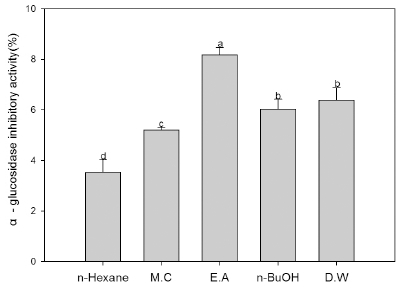
Fig. 2.
Inhibition of α-glucosidase activity using different solvent fractions of Codonopsis lanceolata. Means with the same letter in column are not significantly different at p<0.05 level by Duncan’s multiple range test. The bars represent the standard error. M.C: methylene chloride. E.A: ethyl acetate, n-BuOH: n-butyl alcohol, D.W: distilled water.Pancreatic lipase inhibition
The inhibitory effect of different solvent fractions from C. lanceolata against pancreatic lipase was investigated that are shown in Fig. 3. The pancreatic lipase inhibitory activity of C. lanceolata was found to become a higher effect in ethyl acetate fraction. Inhibition of lipase activity of the ethyl acetate fraction and n-hexane fraction showed a relatively high, while the extract of DW fraction showed the lowest level at given experiment concentration. Obesity is an increasingly serious global problem, not only for the harm it causes in its own right, but also due to the associated health threats, especially type 2 diabetes, systemic hypertension, cardiovascular disease, certain cancers, asthma, and sleep apnea (Kopelman, 2000; Finer, 2006). Many edible plants present an exciting opportunity for the development of newer therapeutics for biologically active antihyperlipidaemic agents from natural resources, especially the reduction of fat digestion and absorption (Kurihara et al., 2006; Zhang et al., 2008; Moller et al., 2009). Pancreatic lipase inhibitors which help to limit intestinal fat absorption at the initial stage, have been proved as useful medications for the treatment of hyperlipidaemia and a great promise as anti-obesity agents (Sharma et al., 2005). The mechanism of lipase inhibitors is to block the absorption of fat by inhibiting lipase in the gastrointestinal tract (Davidson, et al., 1999). Consequently, the root of C. lanceolata in this study may have great potential as dietary supplements or nutraceutical foods for lipase inhibitory effect with antihyperlipidaemic properties.