

Common buckwheat (Fagopyrum esculentum Moench.) is an important crop in some regions of the world. Buckwheat belongs to Polygonaceae family and is believed to have originated from China, and is taxonomically distant from Gramineae to which cereals such as rice, wheat, and maize belong (Tahir and Farook, 1988). Genus Fagopyrum has many species distributed in different parts of the world (Ohnishi, 1998). However, buckwheat has many chemical and morphological characteristics in common buckwheat with cereals, and thus is usually classified as a cereal. Common buckwheat is essentially self-incompatible and posses two main types of flowers: pin and thrum, borne on different plants; these features make the genetic improvement of buckwheat very difficult. Due to allogamy, the individuals in a population are highly heterogenic and the storage proteins show high polymorphism (Dolinsek, 1980; Shevchuk, 1988; Nishiyama et al., 1991).
Buckwheat is consumed mainly in the form of noodles and dumplings. The protein content in common buckwheat varies from 7 to 21%, depending on the cultivar and environmental factors during growth. Most currently grown cultivars yield seeds with 11~15% protein on a whole seed basis. The major protein fractions are globulins which represent almost one-half of all the proteins and consist of 12 to 13 subunits with molecular weights between 17,000 and 57,000. Other known buckwheat protein fractions include albumins and prolamins. The albumin fraction, with a molecular weight of 7,000~8,000, consists of at least 12 proteins. Parliament has been fractionated into at least two peaks by gel filtration and into three major and several minor components by SDS-PAGE. The high nutritive value of the grains and the presence of high content of rutin in foliage makes buckwheat important for human consumption and undoubtedly, is a very important crop in the Japanese diet.
Compared to other cereals, buckwheat grains are rich in essential amino acids such as lysine, threonine, and tryptophan. Although demand for buckwheat is increasing year and year, the year is relatively low and unstable. The complex of proteins present in the seeds of various buckwheat cultivars has proven difficult to resolve analytically, to interrelate functionally and genetically, and to name in a useful manner. Given a heterostyly gene complex (duplication), and the existence of numerous these varieties, the core analytical problem appears to be that of identifying families of related genes and their protein products. Identification of species-specific proteins, if they exist, should be helpful in studies of genetics, taxonomy and evolutionary relationships among buckwheat varieties. Recent developments of proteomics technique have facilitated to study these proteins in a greater depth. Twodimensional gel electrophoresis, combined with very sensitive silver staining, facilitates the analysis of a great number of protein spots on a single gel.
The mechanism of seed storage protein accumulation in buck wheat seeds using SDS-PAGE is reported by Nair and Adachi, (1999). The pointed that, electrophoretic analyses of buckwheat seed proteins during initial seed maturation stage revealed a high molecular mass (69 kDa) protein band. The 24 kDa subunit was reported as allergic protein subunit by Urisu et al. (1994). The intravarietal heterogeneity for different subunits was also reported by Svetek (1994). The major class of buckwheat protein was reported as 13s globulin by Svetlana et al. (1996). Recently, in the field of plant proteomics, plant biology has been increasing interest in the proteins expressed by the genome, the proteome (Pandy and Mann, 2000).
In this context, two-dimensional gel electrophoresis is a powerful tool to analyze complex protein mixtures by separating the proteins according to their isoelectric point (pl) and their molecular mass, as first described by 0`Farrell (1975). Although these technologies had been improved by the development of the better carrier ampholyte production, acrylamide polymers for isoelectric focusing, and experimental instrumentation, probably, the single most important advance in isoelectric focusing for proteome analysis was the development of immobilized pH gradient (IPG) technology (Görg et al., 1992). Also, fingerprint is obtained with the aid of high-resolution techniques such as two-dimensional electrophoresis.
To perform of proteome analysis is very important 2-DE techniques that a number of proteins are separated by mainly twodimensional electrophoresis (2-DE) (Woo et al., 2002). Up to now, we have a problem of seed storage proteins separation in buckwheat. Therefore, seed subunit separations are also considered to play important roles in buckwheat breeding and biochemical characterization. We have currently developed apparatus and methods of two-dimensional electrophoresis techniques.
Results and Discussion
SDS-PAGE analysis
The seeds of common Buckwheat were used in this experiment. Electrophoresis was carried out on an SDS-PAGE (sodium dodecyl sulphate-poly acrylamide gel electrophoresis) discontinuous system as described by Laemmli (1970) with few modifications. The proteins were separated on a 12% acrylamide (30%T, 2.5%C) and 10% SDS. The electrophoretic buffer contained 0.025M Tris, 0.192M glycine, pH8.3, 0.1% w/v SDS. The electrophoresis was carried out for about 3h at constant current of 25mA. The electrophoresis system used were Atto, Japan. The gels were stained in 0.25% Coomassie Brilliant Blue R-250 in methanol: water : acetic acid (5:5:1) for 10mts, and then destained using methanol 25% and acetic acid 7.5%. The gels were dried on a gel drying processor (Atto, Japan) at constant temperature of 60°C for 3h.
Two-dimensional gel electrophoresis (2-DE)
Soluble proteins of whole seed storage were examined by two-dimensional gel electrophoresis according to the protocol of O`Farrell (1975). The isoelectric-focusing (IEF) gel solution contained 48.6g urea, 2% ampholytes (pH 3.5-10), 11.8 ml acrylamide/bis solution (29.2%(w/v) acrylamide, 0.8% (w/v) N`-bis-methylene-acrylamide), 20.3ml 10%(v/v) Triton X-100, 4.5 ml Bio-Lyte 5/7, 0.5 ml Bio-Lyte 3/10 and 28.8 ml distilled water. For preparation of the IEF gel, 1.0 ml IEF gel solution was degassed for 15 min, and 1 μl of N,N,N`,N`-tetramethylethylenediamine (TEMED) and 1.3 μl of freshly prepared ammonium persulfate were added. Then, the gel solution was loaded into the gel tube (3.5 mm ø X 13~27 cm). Solid urea was added to the protein sample (100 to 150 μg protein in about 30 μl) until saturated. To a 5 μl sample was added 1 μl IEF solution containing 0.1 ml 10% (w/v) SDS, 0.02 ml Bio-Lyte 3/10, 0.1 ml 2-mercaptoethanol, and 0.2 ml Triton X-100. The upper and lower reservoirs were filled with 0.1N NaOH and 0.06% phosphoric acid, respectively. After the sample solution was loaded, the gels were run at 200V for 1 h, 400V for 30 min, 600 16 h, respectively, at 9°C. Then the gels were removed from the gel tubes, and equilibrated in 3 ml of reducing SDS buffer (62.5 mM Tris-HCl, pH 6.8, 10% (v/v) glycerol, 5% (v/v) 2-mercaptoethanol, 2% (w/v) SDS, and 0.0125 %(w/v) bromophenol blue (BPB) by shaking for 30 min at room temperature.
The second dimension of gel electrophoresis was carried out using a separation gel of 15% acrylamide and a stacking gel of 5% acrylamide according to the procedure of Laemmli (1970). The gels were run with a constant current at 15mM/gel and 4°C until the BPB reached the bottom of the gel. Preparative gels were stained with Silver. The isoelectric point and relative molecular weight of each protein were determinated using molecular weight marker protein kits (Pharmacia LKB, Uppsala, Sweden).
The new instrument used in this study is simple and allows to easily separate with IEF tube gels in adoption 27 cm length (Fig. 1). Using this instrument, the highly reproducible IEF can be formed in polyacrilamide gels (Fig. 3). Actually, seed storage proteins were separate by 2-DE in the first dimension using IEF tube gels and detected by silver staining.

Results and Discussion
Protein extraction and separation
Extraction of common buckwheat seed proteins is known to be difficult due to reasons; the presence of less ionizable side chains in the major class of common buckwheat protein and the presence of wide range proteins, from very low abundant functional proteins to very high abundant proteins. A combination of urea-mercaptoethanol and NP-40 is a popular choice of solvent used for extraction of common buckwheat seed proteins. In our study, we found that the urea-based solvent is suitable for the extraction of storage proteins but not suitable for low abundant functional proteins. The improved extraction of common buckwheat protein by ureabased solvent could be explained by the fact that the urea has similar molecules as the polypeptide backbone of the globulin protein resulting is very small enthalpy. Alternatively, in our study, good extraction of soluble and low abundant functional proteins was achieved by using 0.1% SDS (Fig. 2), a negatively charged detergent reported to be useful for the extraction of membrane proteins. In our study, we used lysis buffer extracted proteins to compare 2-DE apparatus proteome maps of buckwheat and 0.1% SDS in Tris-HCL extracted proteins to compare nonstorage functional proteins expression in cultivated common buckwheat. To avoid any systematic error from the electrophoresis and staining procedures, we ran two rod gels in one large size SDS-PAGE using a recently introduced electrophoresis apparatus (Fig. 2).

Two-dimensional electrophoresis (2-DE) as a tool for the efficient isolation of seed storage proteins from common buckwheat
Development of a protein isolation protocol that is compatible with isoelecric focusing was integral to developing reproducible 2-D gel proteome reference maps of common buckwheat seed storage protein. Preliminary experiments revealed that protein extracted from common buckwheat seed, in native or denaturing conditions, followed by acetone precipitation of protein prior to a final resuspension in IEF extraction media resulted in highly viscous protein sample. Analysis of this protein fraction by 2-D electrophoresis (50 μl protein loading) resulted in substantial horizontal streaking which suggested contamination with polysaccharides, nucleic acids or other non-protein macromolecules. Insertion of a phenol extraction step immediately after seed crushing effectively partitioned the protein sample from these interfering contaminants as observed by reduced viscosity in the final sample and minimal horizontal streaking on 2-D gels at greater than 50 μ l protein loadings.
Using this method, the highly reproducible isoelectric focusing (IEF) can be obtained from polyacrylamide gels, and IEF from the polyacrylamide gel at all the possible pH range (5.0~8.0) was more easily separated than IPG (immobilized pH gradient) gels.
The polyacrylamide gels in the first dimension in 2-DE was used to separate and identify a number of common buckwheat seed proteins in the proteome analysis. In this new apparatus using 2-DE, 27 cm in length of plate coated with polyacrylamide gel was used and the experiment was further investigated under the various conditions (Fig. 1).
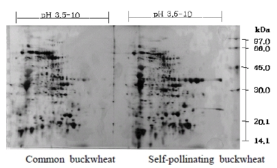
To separate of seed storage protein (50 μl) from three buckwheat species, common buckwheat, self-pollinating buckwheat and tatary buckwheat, resolved by two-dimensional electrophoresis revealed strong similarities in the overall distribution and polypeptide expression pattern (Fig. 2 and 3). The high level of similarity indicated reproducibility in the protein extraction and electrophoretic separation methods. Although the loading of high quantities of protein was necessary to observe lower abundance proteins this frequently resulted in the coalescing of abundant isoelectric protein series (Fig. 2 and 3). Although this broad dynamic range is the result of a few seed storage proteins it is problematic for seed proteome investigations because the preponderance of these proteins masks the detection of other proteins. Since the amount of protein that can be loaded onto an SDS-PAGE is limited to 100 μl-200 μl, based on our experience, in-depth buckwheat seed proteome investigations using 2-D electrophoresis will likely require pre-fractionation steps or narrow range SDS-PAGE for isoelectric focusing. Narrow pH range SDS-PAGE allow for greater protein loads to increase the number of detectable spots.