

Phytolacca americana L. is a perennial plant belonging to the family Phytolaccaceae and is a native plant of North America. This plant was introduced to Korea from the native country, now found in both grassland and forest. P. americana have a toxic in all parts of stem, leaf, fruit, and root. Especially, the root has the highest toxic, but ripe fruit is known to have relatively low toxicity. In addition, its root has been used as a traditional crude diuretic drug, therapeutic purposes of rheumatoid arthritis and swelling in spite of having a strong toxicity. And the pigment extracted from the fruit has been used as additive material of foods and coloring material on the nails of women. P. americana poisoning is a benign plant intoxication that causes gastrointestinal symptoms, including abdominal cramps, vomiting, diarrhea, and gastrointestinal bleeding (Kim et al., 2008). The biological activities of P. americana have been investigated by many researchers with their substance research (Shao et al., 1999; Bylka and Matlawska, 2001; Wang et al., 2008). Recently, plant and plant-derived products are treated a part of the healthcare system by applying the bioactive phytochemicals. Medicinal plants, including P. americana are good sources of antimicrobial agents. Many infectious diseases have been known to be treated with herbal extracts. The clinical efficacy of many existing antibiotics is being threatened by the emergence of multidrug-resistant pathogens (Goveas and Abraham, 2013). The evaluation of antimicrobial property of P. americana is of great interest and importance. Antioxidant compounds in food play an important role as a health protecting factor. Scientific evidence suggests that antioxidants reduce the risk for chronic diseases, including cancer and heart disease. Most of the antioxidant compounds in a typical diet are derived from plant sources and belong to various classes of compounds with a wide variety of physical and chemical properties. The main characteristic of an antioxidant is its ability to trap free radicals. Highly reactive free radicals and oxygen species are present in biological systems from a wide variety of sources. These free radicals may oxidize nucleic acids, proteins, lipids or DNA and can initiate degenerative disease. Antioxidant compounds like phenolic acids, polyphenols and flavonoids scavenge free radicals such as peroxide, hydroperoxide or lipid peroxyl and thus inhibit the oxidative mechanisms that lead to degenerative diseases (Prakash et al., 2001; Boo et al., 2012). In response to the increased production of oxygen radicals the capacity of the antioxidant defense system is increased, but in most situations the response is moderate (Foyer et al., 1994). Tyrosinase is known to be a key enzyme for melanin biosynthesis in plants, microorganisms and mammalian cells. Many tyrosinase inhibitors have been tested on cosmetics and pharmaceuticals as a way of preventing overproduction of melanin in the epidermal layers (Shimizu et al., 2003). Many reports have been found about the tyrosinase inhibitory activity of medicinal plants. P. americana is well known to affect various pharmacological effects for human health. The present study was focused to evaluate of antimicrobial effect, antioxidant and tyrosinase inhibitory activities of the different parts in P. americana.
MATERIALS AND METHODS
Plant material
The sample of Phytolacca americana was collected from Jogye mountain area of Jeonnam province in 2014. Plant part materials (root, stem, leaf and fruit) were freeze-dried, and powdered in a grinder. Each sample powder was stored at -20℃ for experiments. The plant powder was extracted with methanol for 24 hrs. The extracts were freeze-dried after evaporating the solvent under vacuum at temperature below 50℃.
Antimicrobial screening test Strains and media
Strains and media
For the purpose of antimicrobial evaluation, two positive bacteria, two negative bacteria, and one fungus were employed. These microorganisms were purchased from KCTC (Korean Collection for Type Culture, Daejeon, Korea) and KCCM (Korean Culture Center of Microorganisms, Seoul, Korea), and cultured in nutrient agar. Table 1 presents the test microorganisms and culture media.
Table 1.
List of strains and cultivation conditions used for the screening of antimicrobial activity.
Agar diffusion method
The effects of P. americana extracts on five microorganisms (Escherichia coli, Malassezia furfur, Staphylococcus epidermidis, Vibrio parahaemolyticus and Listeria monocytogenes) were evaluated using the agar diffusion method. Inocula of approximately 10 7 CFU were inoculated onto the surface of pre-dried agar. Sterile 8-mm filter paper discs were placed on the plates and impregnated with 40 μL of sample extract. After allowing 1 h at room temperature for the extracts to facilitate diffusion across the surface, the plates were incubated at 37℃ for 24 h for the bacteria. The antimicrobial activity was measured as the size of the clear zone of growth inhibition. The kanamycin was used as the control.
Assay of DPPH radical scavenging rate
100 μL of various concentrations (100, 250, 500, 1000, 2500, 5000 and 10000 mg L-1 ) of extracts of P. americana were added to 900 μL of 100% methanol containing 100 μM DPPH, and the reaction mixture was shaken for 5 min in the slight vortex. Leaving room temperature for 30 min under darkness, the absorbance of DPPH was determined by spectrophotometer at 517 nm. The DPPH radical scavenging activity was calculated according to the following equation: Scavenging effect on DPPH radical (%) = [(A-B)/A]x100, Where A is the absorbance at 517 nm without pigment compositions and B is the change in absorbance at 517 nm with pigment compositions incubation (Brand-Williams et al., 1995).
Assay of Nitrite scavenging rate
The nitrite scavenging activity (NSA) was determined according to a method using Griess reagent (Kato et al., 1987). First, 40 μL of each sample was mixed with 20 μL of 1 mM nitrite sodium. Then the mixture was added to 140 μL of 0.2 M citrate buffer (pH 1.2, 4.2, or 6.0). The final volume of each sample wad adjusted to 200 μL. After, the mixtures had been incubated for 1 h at 37℃, and added to 1000 μL of 2% acetic acid and 80 μL of Griess reagent (1% sulfanilic acid and 1% naphthylamine in a methanol solution containing 30% acetic acid). After vigorous mixing with a vortex, the mixture was placed at room temperature for 15 min, and absorbance was measured at 520 nm. The nitrite scavenging activity was determined based on the following formula:
Where A is the absorbance of the mixture sample during a reation with 1 mM NaNO2 after a 1 h reaction, B is the absorbance of a mixture of distilled water and 1 mM NaNO2 after a 1 h reaction and C is the absorbance of the sample.
(2) Assay of ABTS radical scavenging rate
The spectrophotomeric analysis of ABTS (2,2'-azinbis-(3- ethyl-benzothiazoline-6-sulfonic acid) radical cation (ABTS •+ ) scavenging activity of Lactuca indica was determined according to the method described previously (Re et al., 1999) Re et al., 1999). 7 mM ABTS solution with 2.45 mM potassium persulfate was mixed, and the mixture was incubated in the dark at room temperature for 15 hours, and then was diluted to the absorbance 0.7 at 734 nm. 50 μL of each sample prepared in different concentrations with 950 μL diluted solution was added, and was shaken for 10 seconds by vortex mixer, and then was reacted for 5 min at room temperature, and the absorbance was read at 734 nm using a spectrophotometer (Biochrom Co., England). The ABTS •+ scavenging activity showed as RAEAC (relative ascorbic acid equivalent antioxidant capacity), was calculated by the following equation:
$$\mathrm{RAEAC}=\frac{\mathrm{Caa}}{\mathrm{ΔAaa}}\times\frac{\mathrm{ΔAs}}{\mathrm{Cs}}$$ΔAaa : change of the absorbance after addition of ascorbic acid
Caa : concentration of ascorbic acid
ΔAs : change of the absorbance after addition of sample solution
Cs : concentration of sample
Assay of the tyrosinase inhibitory activity
The assay was performed with slight modifications of Flurkey et al. (2008) method. 0.2 mL of mushroom tyrosinase solution (100 units/mL), 0.6 mL of a 10 mM 3,4-Dihydroxy-L-phenylalanine (DOPA) solution in 0.1 M potassium phosphate buffer (pH 6.8) and 0.2 mL of dimethyl sulfoxide (DMSO) with a sample were mixed. The assay mixture was incubated at 37℃ for 20 min. Following incubation, the amount of dopachrome produced in the reaction mixture was determined spectrophotometrically at 475 nm. The percentage of inhibition of tyrosinase activity was calculated as inhibition (%) = (1 – A/B) x 100, where A represents the absorbance of control solution, and B represents the absorbance of test substance solution.
Data analysis
All experiments were conducted for three to five independent replicates. The data are expressed in terms of mean and standard error. Data were performed using the procedures of the Statistical Analysis System (SAS version 9.1). ANOVA procedure followed by Duncan Multiple Range Test was used to determine the significant difference at the P < 0.05 level.
RESULTS AND DISCUSSION
Antimicrobial activity
The comparative analysis results of the antimicrobial activity of selected microbes (Escherichia coli, Malassezia furfur, Staphylococcus epidermidis, Vibrio parahaemolyticus and Listeria monocytogenes) using the agar diffusion test in the extracts from P. americana are shown in Table 2 and Fig. 1. The antimicrobial activity of P. americana was relatively high in Malassezia furfur known as a skin fungi and Vibrio parahaemolyticus compared to Escherichia coli and Staphylococcus epidermidis. However, the antimicrobial activity in Vibrio parahaemolyticus did not show at all parts of P. americana. The antimicrobial activity in different plant parts was relatively the highest in the leaf. The antimicrobial activity of P. americana is thought to look different depending on the bacterial strains and the different plant parts. Overall, the extracts of P. americana were more antimicrobial activity on fungus and gram positive bacteria than gram negative bacteria. Currently a wide range of natural substances known to have antimicrobial activity (Chan et al., 2008; Goveas and Abraham, 2013; Modaressi et al., 2013), but there is little research on related to the antimicrobial efficacy and the application of P. americana. It is not surprising that there are differences in the antimicrobial effects of plant species, due to the phytochemical properties and differences among species. Some of plant extracts may have contained antibacterial constituents, just not in sufficient concentrations so as to be effective. It is also possible that the active chemical constituents were not soluble in methanol or water (Stainer et al., 1986; Parekh and Chanda, 2007). The potential for developing antimicrobials from higher plants appears rewarding as it will lead to the development of a phytomedicine to act against microbes. Plant-based antimicrobials have enormous therapeutic potential as they can serve the purpose with lesser side effects that are often associated with synthetic antimicrobials (Iwu et al., 1999). Further research is necessary to determine the identity of the antibacterial compounds from within P. americana and also to determine their full spectrum of efficacy.
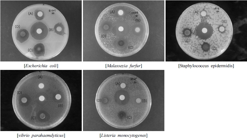
Fig. 1.
Inhibition activity of methanol extracts in different parts from Phytolacca Americana against five microorganisms in paper disc diffusion assay. A:Root, B:Stem, C:Leaf, D:Fruit.Table 2.
Inhibitory activity against the microorganisms by agar diffusion method of methanol extracts in different parts from Phytolacca Americana.
DPPH radical scavenging activity
Free radical scavenging is one of the most important accepted mechanisms for antioxidant activity. DPPH stable free radical scavenging method can be used to evaluate the antioxidant activity of a extracts in a short time (Tiwari, 2001; Hosseinimehr et al., 2007). The values of the 50% inhibition concentration (IC50) of leaf, fruit, stem and root extracts of P. americana for the DPPH radical were 10991.76 mg L-1 , 21915.45 mg L-1 , 41496.50 mg L-1 and 46264.22 mg L-1 , respectively (Table 3). As it is shown in Table 3, leaf demonstrated the highest DPPH scavenging capacity compared to other plant parts. The investigation of the antioxidant activity of natural substances is based on the measuring of the electron donor capacity of DPPH with the ability to inhibit the oxidation by donating electrons in free radicals causing this lipid peroxidation. Active oxygen caused by in vivo metabolism removed by the body's antioxidant system, but excessive free radicals induced stress, causing the lipid peroxidation by combining with unsaturated fatty acids in the cell membrane, and brought intracellular structural and functional damage. Looking at the results, the DPPH scavenging capacity of the extracts of P. americana showed that the increase was proportional to the concentration. Cells are oxidized and damaged by the free radical, depending on the growth of cells. It has been reported that phenolic compounds have antioxidant capacity to inhibit the oxidation by donating electrons to the free radical due to a strong reduction (Sanchez et al., 2007; Saija et al., 1998). In this study, the DPPH radical scavenging activity appeared to concentration dependent, and depending on the different plant parts, there were significant differences in the results.
Table 3.
DPPH radical scavenging activities of Phytolacca americana at different plant parts.
| Plant | Plant Part | DPPH radical scavenging activity, % of control | ||||||
|---|---|---|---|---|---|---|---|---|
| Concentration (mg/L) | ||||||||
| 500 | 1000 | 2500 | 5000 | 10000 | 20000 | IC50 | ||
| Phytolacca americana | ||||||||
| Root | 8.54±0.20c | 9.42±0.14c | 11.27±0.12c | 13.09±0.17c | 16.53±0.10c | 27.10±0.40d | 46264.22 | |
| Stem | 9.39±0.21b | 10.46±0.09b | 11.36±0.23bc | 12.61±0.21c | 16.28±0.15c | 29.34±0.77c | 41496.50 | |
| Leaf | 11.78±0.17a | 14.18±0.19a | 24.15±0.15a | 29.38±0.23a | 49.78±1.34a | 78.45±0.89a | 10991.76 | |
| Fruit | 8.72±0.29bc | 9.13±0.14d | 11.85±0.06b | 13.96±0.24b | 24.06±0.14b | 48.27±0.72b | 21915.45 | |
ABTS radical scavenging activity
In order to evaluate the radical scavenging activities of methanol extracts of different parts from P. americana, ABTS assays were performed. The results of the ABTS radical scavenging activity were shown in Table 4. When the different parts from P. americana were treated with various concentrations (1000, 2500, 5000, 10000 and 20000 mg L-1 ) of methanol extracts, the ABTS radical scavenging activity was progressively increased in a dose-dependent manner. However, methanol extracts at 20 mg/mL from leaf showed a manifested radical scavenging activity (67.34%) compare to that of methanol extract from fruit (40.72%), stem (38.69%) and root extract 29.57%). The IC50 values (50% inhibition concentration) was observed from the leaf extract (IC50 of 13546.67 mg L-1 ), followed by fruit (IC50 of 25633.44 mg L-1 ), stem (IC50 of 26430.00 mg L-1 ) and root (IC50 of 36352.75 mg L-1 ). In the present evaluation, both the DPPH radical scavenging activity and ABTS radical scavenging activity have been increased with the higher concentration of methanol extract. Leaf extract of P. americana exhibited the highest activity both ABTS radical scavenging activity and DPPH radical scavenging activity. All samples of P. americana organ proved that ABTS radical scavenging activity were dose dependent. It revealed that the studied plants showed a dose dependent ABTS radical scavenging activity (Lee et al., 2006).
Table 4.
ABTS radical scavenging activities of Phytolacca americana at different plant parts.
| Plant | Plant Part | ABTS radical scavenging activity, % of control | |||||
|---|---|---|---|---|---|---|---|
| Concentration (mg/L) | |||||||
| 1000 | 2500 | 5000 | 10000 | 20000 | IC50 | ||
| Phytolacca americana | |||||||
| Root | 7.76±0.13d | 9.53±0.11d | 10.65±0.15d | 19.99±0.23d | 29.57±0.28d | 36352.75 | |
| Stem | 10.00±0.11b | 12.23±0.07c | 20.41±0.23b | 28.54±0.15b | 38.69±0.47c | 26430.00 | |
| Leaf | 13.54±0.19a | 21.99±0.15a | 28.36±0.33a | 28.36±0.33a | 67.34±0.14a | 13546.67 | |
| Fruit | 9.11±0.10c | 13.68±0.16b | 17.04±0.27c | 27.33±0.36c | 40.72±0.43b | 25633.44 | |
Nitrite scavenging activity
Nitrite ions in the acidic environment of the stomach induce mutagenic and cell-damaging reactions (Kato and Puck, 1971). Exposure to excess nitrite from the diet is implicated as a potential etiological factor in the development of stomach and colorectal cancers (Lee et al., 2006). Nitrite reacts with second and third grade amines to form nitrosamine in protein-rich foods, medicines, and residual pesticides. It is also present in large quantities in meat and both leafy and root vegetables. Nitrosamine is converted to diazoalkane (alkane nucleic acid), proteins, and intracellular components, which can increase the risk for cancer (Choi et al., 2008). In order to investigate the nitrite scavenging activity of extracts of different parts from P. americana, various acidic conditions were tested. The results of the nitrite scavenging activity were shown in Table 5. The nitrite scavenging activities were affected by the changes in pH. The nitrite scavenging activity was decreased when the pH was changed from pH 1.2 to pH 6.0. The highest nitrite scavenging activity was exhibited from the methanol extract of fruit (71.31%), followed by root (63.30%), stem (59.00%) and leaf (32.87%) at pH 1.2. The fact that the nitrite scavenging activity was high at pH 1.2 suggests that nitrosamine production can be inhibited in vivo (Choi et al., 2008). These results were consistent with other findings that had the highest the nitrite scavenging at pH of 1.2 in fermented pine extract (Hong et al., 2004) and extracts from different parts of citron (Shin et al., 2005). However, the nitrite scavenging activity at pH of 6.0 was not almost detected.
Tyrosinase inhibitory activity
Tyrosinase is the key enzyme in the first stage of the melanogenesis pathway, catalyzing the conversion of L-tyrosine into L-dopaquinone (Marmol et al., 1993).Tyrosinase is also one of the key enzymes responsible for controlling the insect molting process (Nevil, 1975), and could therefore be used as an insecticide for the control of pests and insects. Tyrosinase inhibitors have become increasingly important as cosmetic and medicinal products, primarily to control melanin pigmentation (Khanom et al., 2000). The results of the tyrosinase inhibitory activity of methanol extracts from the different plant parts were shown in Fig. 2. All organs of P. americana showed tyrosinase inhibitory activity. The highest activity was found in the stem, and followed by root, leaf, and fruit in order. These tyrosinase inhibitory activity was progressively increased in a concentration-dependent manner. In the present study, we confirmed that the extract of P. americana showed potential tyrosinase inhibitory activity of the methanol extracts of different organ from P. americana. Therefore, the natural material from P. americana broadens the possible use of tyrosinase inhibitors as food additives, in addition to insect control agents and whitening agents.