

Many researches have shown that traditional medicinal plants have in vitro mutagenic or toxic and carcinogenic properties, thus it is important to explore the medicinal plants for their cytotoxicity. The cytotoxicity evaluation of plants is a major subject in pharmaceutical studies, particularly in the area of cancer research (Cuyacot et al., 2014). However, there is a scarcity of data on the safety and tolerability of Platycodon grandiflorum when used as a health care materials. Medicinal plants including P. grandiflorum are good sources of antimicrobial agents. Many infectious diseases have been known to be treated with herbal extracts. The clinical efficacy of many existing antibiotics is being threatened by the emergence of multidrug-resistant pathogens (Goveas and Abraham, 2013). The evaluation of antimicrobial property of tetraploid P. grandiflorum is of great interest and importance. Most of the antioxidant compounds in a typical diet are derived from plant sources and belong to various classes of compounds with a wide variety of physical and chemical properties. The main characteristic of an antioxidant is its ability to eliminate free radicals. Highly reactive free radicals and oxygen species are present in biological systems from a wide variety of sources. The plants have antioxidant enzymes such as superoxide dismutase (SOD), catalase (CAT), peroxidase (POD), and ascorbate peroxidase (APX) against ROS (reactive oxygen species) (Zhou et al., 2005). Both enzymatic and nonenzymatic antioxidant systems are present in plants. Superoxide radicals are detoxified by SOD and hydrogen peroxide is destroyed by CAT and different kinds of peroxidases (Kang and Saltveit, 2002). A major hydrogen peroxide-detoxifying system in plant is the ascorbate-glutathione cycle that includes APX and glutathione reductase (GR) (Asada, 1994). Ascorbate peroxidase, catalase and peroxidase, together with low-molecular mass scavengers such as ascorbate, glutathione and proline, act as the main defense against ROS produced in various parts of plant cells (Apel and Hirt, 2004). The induction of ROS-scavenging enzymes, such as SOD, POXs and CAT, is the most common mechanism for detoxifying ROS synthesized during stress responses (Wojtaszek, 1997; Mittler, 2002). P. grandiflorum is a perennial flowering plant belonging to the family Campanulaceae and is grown commercially in East Asia. Roots of P. grandiflorum have been used as a traditional oriental medicine and food for bronchial asthma, hepatic fibrosis, bone disorders (Lee et al., 2004; Choi et al., 2009; Jeong et al., 2010; Lee, 1973), hypercholesterolemia and hyperlipidemia (Kim et al., 1995). Roots of P. grandiflorum containing triterpenoid saponin, inulin, phytosterin, platycidinin, proteins, lipids, carbohydrates, iron, and fibers are similar to the ginseng roots and are being cultivated for food or medicine material. Recent studies indicate that platycodins are one of the most essential functional components in P. grandiflorum in terms of the inhibition of pancreatic lipase (Zhao and Kim, 2004), cholesterol lowering, and antiobesity effects (Zhao et al., 2006). P. grandiflorum is well known to affect various pharmacological effects for human health and its consumption is increasing. In order to develop functional products using the physiological functionality, P. grandiflorum is needed a mass production of natural materials and the breeding of superior varieties. The creating of giant P. grandiflorum by the polyploidy breeding method can maximize its effects. The polyploidy breeding method in plants is a way to increase radically the emergence of new useful traits and the quantity by polyploidy obtained through quantitative doubling of the genome, which is a set of chromosomes. Polyploids, although frequently encounter low seed setting rates or complete sterility (Lewis, 1980), usually show larger organ size and superior cold tolerance (Kato and Birchler, 2006). For medicinal plants, polyploidy may increase the amounts of secondary metabolites (Thao et al., 2003) which functional compounds accumulate in the vegetative parts such as purple coneflower (Gao et al., 1996). So, polyploidy breeding is an effective approach of germplasm development for medicinal plants. Methods using colchinine for polyploidy induction are common for a wide range of plant species (Luckett, 1989; Ishizaka and Uematsu, 1994; Pinheiro et al., 2000; Petersen et al., 2003; liu et al., 2007). Some reports were presented that the tetraploid induction of P. grandiflorum by colchicine treatment have also been available for breeding (Kim et al., 2003; Wang et al., 2006; Wu et al., 2011). The present study was focused to evaluate the cytotoxicity, antimicrobial and antioxidant enzyme activity of diploid and tetraploid P. grandiflorum.
MATERIALS & METHODS
Plant material
The diploid plant of Platycodon grandiflorum was grown in Geumsan county, Chungcheong province and purchased from local market. Tetraploid plants were provided by the cooperative research laboratory of this study, Chungbuk National University. Root samples were freeze-dried, indoor-dried, hot-air dried, and microwave dried and then ground. Each sample powder was stored at -20℃ for experiments.
Induction of tetraploid
Tetraploid mutants were induced in a similar method to the procedure described by Kim et al. (2003). The colchicine treatment on seedlings were performed at the time of cotyledon emergence. After the treatment, seedlings were washed 3~4 times with distilled water and planted on 12cm pots containing of equal volume of perlite and coarse sand. Survival rate and chromosome numbers were counted 30 days after transplanting. After 4 months, measurement of stomates and morphological characters were made. To induce polyploids in adult plants, growing points were covered with cotton balls and sprayed with concentration of 0.05% colchicines solution 3 times a day for 3 days. The ploidy level of Platycodon grandiflorum was estimated chromosome counting of root tips from obtained mutants by morphological characteristics.
Cytotoxicity measurement by the MTT assay
The cytotoxicity of Platycodon grandiflorum sample was assayed using human cancer cell lines, HeLa for human metrocarcinoma, Calu-6 for human pulmonary carcinoma, MCF-7 for human breast adenocarcinoma, HCT-116 for human colorectal carcinoma, SNU-1066 for human laryngeal squamous cell carcinoma, Hep 3B for human hepatocellular carcinoma and SNU-601 for human gastric carcinoma. The cell lines were purchased from Korea Cell Line Bank (KCLB) for MTT (3-(4,5-dimethylthiazol-2-yl)-2,5-diphenyltetrazolium bromide) assay. The cells plated on 96 well plates at a concentration of 3 x 10⁴ cells/mL. The cells were incubated for 24 hrs in RPMI-1640 medium at 37°C under 5% CO2 in a humidified incubator, and treated with 2 μL of various concentrations (50, 100, 200, 400, and 800 μg mL-1) of extracts. After the incubation for 48 hr, the cells were washed twice with phosphate buffer solution (PBS). MTT solution at 5 mg/mL was dissolved in 1mL of PBS, and 10 μL of it was added to each of the 96 wells. After the reaction for 4 hr, the solution in each well containing media, unbound MTT and dead cells were removed by suction and 100 μL of DMSO was added to each well. The plates were shaken for 15 minutes by plate shaker, and the absorbance was recorded using an ELISA reader (Bio-Rad model 550, USA) at a wavelength 540 nm. The viability of the treatment was determined as percentage of viability compared to untreated cell, and the values were then used to iteratively calculate the concentration of plant extracts required to cause a 50% reduction (IC50) in growth for each cell line.
Antimicrobial screening test on the bronchus disease bacteria
Strains and media
For the purpose of antimicrobial evaluation, 2 g of positive bacteria, and 4 g of negative bacteria were employed. These microorganisms were purchased from the Korean Collection for Type Culture (KCTC, Daejeon, Korea) and cultured in nutrient agar. Table 1 presents the test microorganisms and culture media.
Table 1.
List of strains and cultivation conditions used for screening of antimicrobial activity test.
Agar diffusion method
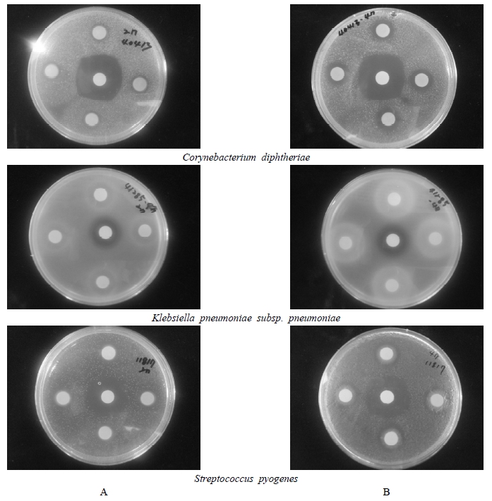
The effects of P. grandiflorum extracts on the bronchus disease bacteria (Corynebacterium diphtheria, Klebsiella pneumoniae subsp. Pneumonia and Streptococcus pyogenes) were evaluated using the agar diffusion method. Inocula of approximately 107 CFU were inoculated onto the surface of pre-dried agar. Sterile 8-mm filter paper discs were placed on the plates and impregnated with 40 μL of sample extract. After allowing 1 h at room temperature for the extracts to facilitate diffusion across the surface, the plates were incubated at 37℃ for 24 h for the bacteria. The antimicrobial activity was measured as the size of the clear zone of growth inhibition. The kanamycin was used as the control.
Assay of antioxidant enzyme on fraction extracts
SOD activity
The superoxide dismutase (SOD) activity was measured using SOD assay Kit-WST purchased from Sigma-Aldrich (Sigma-Aldrich Co., Japan). This assay is based on the colorimetric assay for the measurement of total antioxidant capacity of crude aqueous fractions. The 60 μL of sample solution (sample and blank2) or doubledistilled water (blank1 and blank3) was mixed with 600 μL of WST working solution. For Blank2 and Blank3, 60 μL of dilution buffer was added. Then, 60 μL of enzyme working solution was added to each sample and blank1. The plate was incubated at 37℃ for 20 min, and the OD (Optical density) was determined at 450 nm using a spectrophotometer (Biochrom Co., England). SOD activity (inhibition rate percent) was calculated using the following equation:
SOD activity={[(Ablank1–Ablank3)–(Asample–Ablank2)] /(Ablank 1–Ablank 3)} ×100.
CAT activity
Catalase (CAT) activity was assayed by the method of Mishra et al. (1993). The reaction mixture contained 50 mM potassium phosphate buffer (pH 7.0), 11 mM H2O2, and the crude enzyme extract. The reaction was initiated by addition of H2O2 to the mixture, and enzyme activity was determined by monitoring the decline in absorbance at 240 nm (ε=36 M− 1 cm−1), because of H2O2 consumption.
APX activity
Ascorbate peroxidase (APX) activity was determined by monitoring the decline of absorbance at 290 nm as ascorbate (ε =2.8 mM−1 cm−1) was oxidized, by the method of Chen and Asada (1989). The reaction mixture contained 100 mM potassium phosphate buffer (pH 7.5), 0.5 mM ascorbate, and 0.2 mM H2O2.
POX activity
Peroxidase (POX) activity was determined specifically with guaiacol at 470 nm (ε=26.6mM−1 cm−1), following the method of Egley et al. (1983). The reaction mixture contained 40 mM potassium phosphate buffer (pH 6.9), 1.5 mM guaiacol, and 6.5 mM H2O2 in 1 ml with crude enzyme extract. Control assays in which the enzyme extracts or substrates were replaced by buffer were performed.
RESULTS & DISCUSSION
Cytotoxicity
The cytotoxicity of P. grandiflorum on seven human cancer cell lines were evaluated by the MTT assay. When cells were treated for 48 hrs with various concentrations (50, 100, 200, 400 and 800 μg/mL) of methanol extracts, the rate of cell survival progressively decreased in a dose-dependent manner. Results of the cytotoxicity evaluation against human cancer cell lines from roots of P. grandiflorum are shown in Table 2. Overall, the cytotoxicity of methanol extracts of P. grandiflorum showed significant differences between tetraploid and diploid. However, the cytotoxic effect against human cancer cell was higher in tetraploid than in diploid. The extract of tetraploid at 200 μg/mL exhibited a pronounced cytotoxic effect (20.02%) on HCT-116 cell compare to that of diploid (60.45%). At all extracts concentration, tetraploid samples showed high toxicity and the IC50 (concentration causing 50% cell death) value showed the highest on HCT-116 cell (105.91 μg/mL), and exhibited significant activity against the Hep 3B cell (140.67 μg/mL), SNU-1066 cell (154.01 μg/mL), Hela cell (158.37 μg/mL), SNU-601 cell (182.67 μg/mL), Calu-6 cell (190.42 μg/mL), MCF-7 cell (510.19 μg/mL). The values of IC50 in diploid samples showed the highest on SNU-1066 cell (239.34 μg/mL), followed by 295.17 μg/mL on Hela cell, 309.12 μg/mL on HCT-116 cell, 393.17 μg/mL on Hep 3B cell, 432.25 μg/mL on SNU-601 cell. On the contrary, the extracts on MCF-7 cell and Calu-6 cell exhibited the weakest inhibition on cell viability, having an IC50 value of over 800 μg/mL against MCF-7 cell (831.91 μg/mL) and Calu-6 cell (947.64 μg/mL). The persistency search for new anticancer compounds in plant medicine and traditional foods is a realistic and promising strategy for its prevention. Numerous compounds found in plants with anticancer properties are such as alkaloids, phenylpropanoids, and terpenoids (Kintzios, 2006; Park et al., 2008; Yan-Wei et al., 2009; Vijayarathna and Sasidharan, 2012). Presently there is an increasing interest world wide in herbal medicines accompanied by an increased laboratory investigation into the pharmacological properties of the bioactive ingredients and their ability to treat various diseases (Lobo et al., 2009). It is well known that chemicals and medicinal plant medicines, may produce toxic effects. Based on the results presented in this paper, tetraploid P. grandiflorum can be used as a source of cytotoxic agent.
Table 2.
Cytotoxicity of extracts on seven human cancer cell lines of diploid and tetraploid in Platycodon gradinflorum.
| Plant | Cell line | Cell viability, % of control | |||||
|---|---|---|---|---|---|---|---|
| Concentration (μg/mL) | |||||||
| 50 | 100 | 200 | 400 | 800 | IC50 | ||
| Diploid | SNU-601 | 80.45±2.13c | 78.23±3.31b | 70.77±5.83b | 64.64±2.59c | 11.39±1.55cd | 432.25 |
| SNU-1066 | 104.82±7.14a | 102.32±5.79a | 77.23±8.08ab | 26.99±3.44d | 5.91±0.48d | 239.34 | |
| MCF-7 | 90.39±3.11abc | 82.66±4.83b | 77.01±3.05ab | 75.05±1.06b | 50.34±2.65b | 831.91 | |
| HCT-116 | 75.75±8.81c | 68.80±4.97b | 60.45±5.09b | 34.18±3.96d | 10.50±2.18d | 309.12 | |
| Calu-6 | 106.24±5.29a | 104.65±8.22a | 94.32±6.78a | 89.98±3.14a | 57.60±4.34a | 947.64 | |
| Hep 3B | 99.60±5.39ab | 74.12±8.86b | 70.77±8.59b | 30.50±4.26d | 17.50±1.08c | 393.17 | |
| Hela | 84.43±6.86bc | 70.94±5.90b | 63.88±7.67b | 16.65±2.05e | 6.22±0.35d | 295.17 | |
| Tetraploid | SNU-601 | 100.23±9.51a | 94.40±9.06a | 49.13±5.32b | 16.03±2.34c | 5.71±0.29d | 182.67 |
| SNU-1066 | 102.01±5.29a | 82.53±7.10ab | 31.58±2.74d | 8.21±0.73bc | 7.00±0.39c | 154.01 | |
| MCF-7 | 96.74±5.91ab | 94.86±5.01a | 90.66±7.69a | 55.51±6.11a | 20.21±3.99a | 510.19 | |
| HCT-116 | 68.65±3.23c | 61.05±9.01b | 20.02±2.83d | 4.76±0.67c | 3.55±0.22c | 105.91 | |
| Calu-6 | 79.94±3.42bc | 75.02±8.80ab | 69.56±4.37b | 13.51±2.64b | 3.66±0.21c | 190.42 | |
| Hep 3B | 86.48±6.78abc | 77.29±4.21ab | 22.56±1.39d | 15.11±2.34b | 12.82±2.10b | 140.67 | |
| Hela | 96.05±4.81ab | 91.24±9.83a | 33.95±5.82d | 8.18±1.00bc | 5.88±0.36c | 158.37 | |
Antimicrobial activity
The antimicrobial activity of methanolic extracts of roots of P. grandiflorum was assayed in vitro by agar disc diffusion method against three different bronchus diseases bacterial strains (Corynebacterium diphtheria, Klebsiella pneumoniae subsp. Pneumonia and Streptococcus pyogenes). The comparative analysis results are shown in Fig. 1 and Table 3. Antimicrobial activities of diploid P. grandiflorum were relatively low compared to tetraploid P. grandiflorum on most of bacterial strains. In tetraploid P. grandiflorum, K. pneumoniae showed the clear zone formation (18~19 mm) of growth inhibition, followed the clear zone formation of 13~15 mm on C. diphtheria and S. pyogenes. The antimicrobial activities in diploid P. grandiflorum was the highest on K. pneumonia (14~15 mm), and showed the clear zone formation of 11~12 mm on C. diphtheria and 12~13 mm on S. pyogenes. The antimicrobial activity is thought to look different depending on the bacterial strains and the polyploidy of P. grandiflorum. It was reported that the antimicrobial activities against bronchus diseases bacterial strains of extracts from ethyl ether and petroleum ether of diploid P. grandiflorum were high (Lee et al., 2000). Currently a wide range of natural substances known to have antimicrobial activity, and also active research underway thereto (Chan et al., 2008; Goveas and Abraham, 2013; Modaressi et al., 2013), but few studies related to the antimicrobial efficacy against bronchus diseases and the application of the tetraploid P. grandiflorum has been done. These results indicate that the tetraploid P. grandiflorum can be used for developing antimicrobials which can act against bronchus diseases bacterial strains.
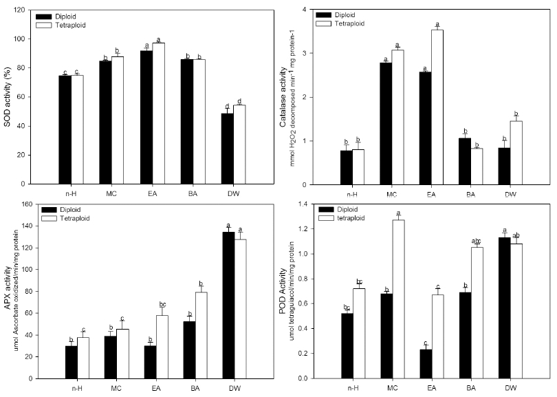
Antioxidant enzyme activity
Freeze-dried samples of P. grandiflorum were extracted in methanol at room temperature and fractionated into five different solvents (hexane, methylene chloride, ethyl acetate, butyl alcohol, and water). This study shows that the plants induced polyploidy mutants can be achieved the activities of SOD, CAT, APX, and POD. The comparative results of the antioxidant enzyme activity in fraction extracts of diploid and tetraploid in P. grandiflorum plants are shown in Fig. 2. The root extract of P. grandiflorum had the highest (97.2%) SOD enzyme activity in ethyl acetate partition layer of tetraploid and while water partition layer of diploid showed the lowest (48.6%) SOD enzyme activity. The activity of CAT showed higher values in the root of tetraploid than in the diploid of P. grandiflorum in all partition layers except butyl alcohol. The activities of APX and POD showed higher values in the root of tetraploid than in the diploid of P. grandiflorum in all fraction solvents except water layer (Fig. 2). Significant roles of POD have been suggested in plant development processes (Gaspar et al., 1985), which was involved in scavenging of H2O2 produced in chloroplasts (Asish and Anath, 2005). The antioxidant enzyme activities differ significantly in different plants. The SOD is one of the enzymes, in vivo, to catalyze the reaction that converts the harmful reduced oxygen formed in cell due to rancidity into hydrogen peroxide; is generated in most aerobic or anaerobic biological organisms; is switched to water and oxygen by the CAT and APX, and loses then its toxicity. APX activity, which is an important component of the antioxidant system, plays a key role in eliminating H2O2 molecules and in the modulation of its steady-state levels in various plant subcellular compartments (Najami et al., 2008). The CAT is also an antioxidant enzyme that protects cells by dispatching of in vivo harmful oxygen and is a typical enzyme that acts to decompose and scavenge the H2O2 together with APX. We consider that relatively high antioxidant enzyme activity in the tetraploid of P. grandiflorum is influenced by providing some stress on the plant under forced polyploidy induction (Boo et al., 2013). The antioxidant enzymes, indicating a high activity to remove harmful free radicals, have the effect of prevention and inhibition of various diseases and aging. In the tetraploid of P. grandiflorum, we can also expect to see these benefits for the next variety of natural foods and cosmetics where the need to apply functional substances may also be required. In conclusion, to use of tetraploid P. grandiflorum in healthy functional foods and medicinal industry with regard to its high antioxidant potentials, it is suggested that complement experiments is required to accomplish on fraction extracts of P. grandiflorum.